MR Imaging of the Gallbladder: A Pictorial Essay - vRad.


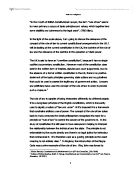
MR images showed a thickened gallbladder wall and intramural cavities, which are hypointense on the T1- weighted images and hyperintense on the T2-weighted images. This thickening was located in the fundus and extended to the body causing important luminal stenosis.

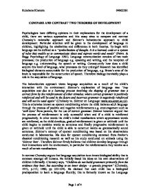
Gallbladder sludge, also known as biliary sand, biliary sediment, or thick bile, is a mixture of particulate matter and bile, normally seen as a liquid-liquid level in the gallbladder on ultrasound, corresponding to the precipitate of bile solutes.

Functional MR cholangiography (fMRC) is an emerging technique that has the potential to provide a comprehensive evaluation for the anatomic and functional assessment of the gallbladder and biliary tree.

In cases of suspected gallbladder disease, sonography is often the first imaging technique because of its relatively low cost and widespread availability (2, 5).Although sonography has a relatively high sensitivity for the detection of tumor at advanced stages, it is limited in the diagnosis of early lesions and is unreliable for staging.

Developed by renowned radiologists in each specialty, STATdx provides comprehensive decision support you can rely on - Hyperplastic Cholecystosis (Adenomyomatosis).

Developed by renowned radiologists in each specialty, STATdx provides comprehensive decision support you can rely on - Gallstones and Sludge.

Imaging Modalities. The modalities used for imaging the biliary tree include US, CT, ERCP, PTC, MRI, and MRCP. Ultrasound. Biliary disease presents clinically with a wide range of manifestations from being asymptomatic to overt signs of biliary obstruction in the form of jaundice or elevated liver function tests.

This condition is notoriously non-specific on imaging on many occasions, particularly in its early stages; gall bladder carcinoma shows numerous features that overlap with a large number of benign conditions, leading to delayed diagnosis and incurable disease. Radiologists should be familiar with its typical and atypical imaging features.
Other posts on the site.

Imaging of common bile duct stones The gallbladder serves as the repository for bile produced in the liver. However, bile within the gallbladder may become supersaturated with cholesterol, leading to crystal precipitation and subsequent gallstone formation.

Sonography is usually the preferred screening study because of its availability, relatively low cost and lack of radiation hazard. Magnetic resonance (MR) imaging can be a valuable complement to US and CT. US is an essential first-line investigation in suspected gallbladder and biliary duct disease. It is usually performed using the highest.

US examination of the gallbladder is a common investigation that has excellent potential in assessing inflammatory and calculus disease. The high-resolution capabilities of US allow clear depiction of gallstone and gallbladder polyps that are often not seen on CT or MR imaging, albeit with the proviso that US has inherent limitations in the difficult patient.

MR imaging of the gallbladder: a pictorial essay. Catalano OA, Sahani DV, et al. Radiographics. 2008 Jan-Feb;28(1):135-55 Best cases from the AFIP: Adenomyomatosis of the gallbladder.
In the case of a duplicated gallbladder, magnetic resonance cholangiopancreatography can be distinguished from a hemangioma (10). In conclusion, the possibility of gallbladder hemangioma, although rare, must be considered if a well-defined sub-epithelial tumor of the gallbladder shows peripheral nodular.

The aim of this pictorial essay is to familiarise the reader with multiple examples of classic aunt Minnie cases specific to the abdomen and pelvis. The emphasis of the cases included is to classic cases in their modern day manifestation that are more representative of current imaging practices. Methods and materials Cases demonstrating all components to constitute an aunt Minnie of the.
Gallbladder adenomyomatosis also known as adenomyoma or adenomyomatous hyperplasia of the gallbladder, is a benign (non-cancerous) condition characterized by epithelial proliferation and hypertrophy of the muscles of the gallbladder wall 1) with an outpouching of the mucosa into or through the thickened muscular layer, i.e., the Rokitansky.

Pathology rising from the gallbladder is a group of entities that every radiologist has had to deal with during its professional carrer. That is why we ilustrate typical and atypical cases in the gallbladder that the radiologist must identify in order to depict abnormalities and the importance of identification in US (classical diagnostic tool) and MR, in which we make special emphasys.